It then reaches the eye’s drainage system, including the trabecular meshwork and a network of canals.

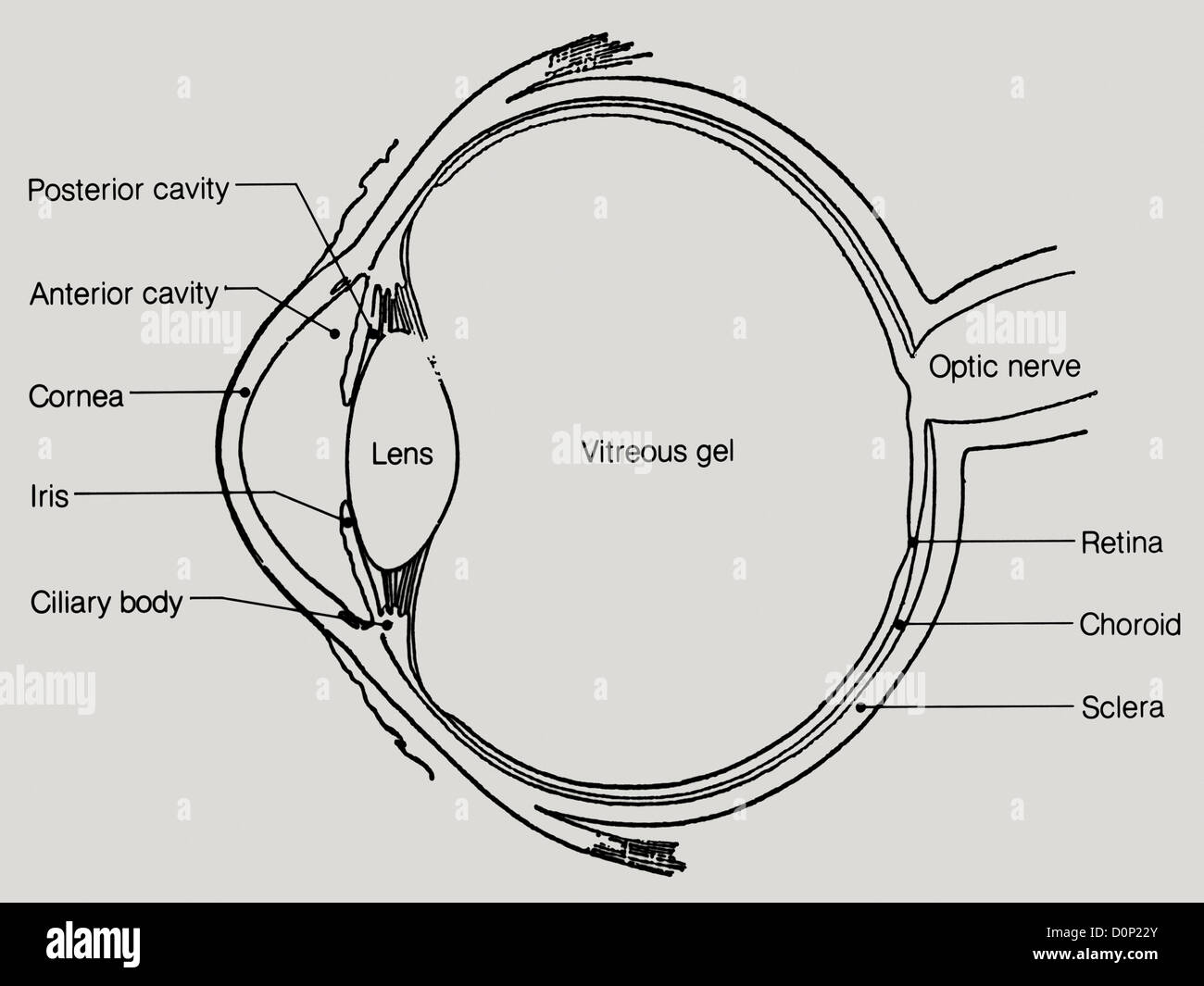
The front part of the eye is filled with a clear fluid (called aqueous humor) made by the ciliary body. The optic disc is the area on the retina where all the nerve fibers come together to become the optic nerve as it leaves the eye to connect to the brain. The retina converts the light images into electrical signals, and the retina’s nerve cells and fibers carry these signals to the brain through the optic nerve. The lens inside our eye focuses this light onto the back of the eye, which is called the retina. At the center of the iris is a hole (covered by the clear cornea) called the pupil, where light enters the eye. The iris, a muscle, is the colored part of the eye that contracts and expands to let light into the eye. At the very front of the eye is a clear surface, like a window, called the cornea that protects the pupil and the iris behind that window. A clear thin layer called the conjunctiva covers the sclera. Marshall, J., Usher, D.: Method for generating a unique and consistent signal pattern for identification of an individual, US Patent No.Understand the Eye to Understand GlaucomaĬovering most of the outside of the eye is a tough white layer called the sclera. Hill, R.B.: Apparatus and method for identifying individuals through their retinal vasculature patterns, US Patent No. Simon, C., Goldstein, I.: A new scientific method of identification. Hill, R.B.: Fovea-centered eye fundus scanner, US Patent No. Snell, R.S., Lemp, M.A.: Clinical Anatomy of the Eye, pp. Online textbook, ∼cse891/Sect601/textbook/5.pdf. In: Biometrics: Personal Identification in Networked Society. 89, 326–333 (2001)ĭaugman, J.: Recognizing persons by their iris patterns.

4,641,349 Feb (1987)ĭaugman, J.: Biometric personal identification system based on iris analysis, US Patent No. Sinauer Associates, Inc., Sunderland (1999)įlom, L., Safir, A.: Iris recognition system, US Patent No.

Oyster, C.W.: The Human Eye: Structure and Function, pp. (eds.): Gray’s Anatomy, 35th British edn. Academic Press, Orlando (1984)īorn, A.J., Tripathi, R.C., Tripathi, B.J.: Wolff’s Anatomy of the Eye and Orbit, 8th edn. Pevsner, J.: Leonardo da Vinci’s contributions to neuroscience.


 0 kommentar(er)
